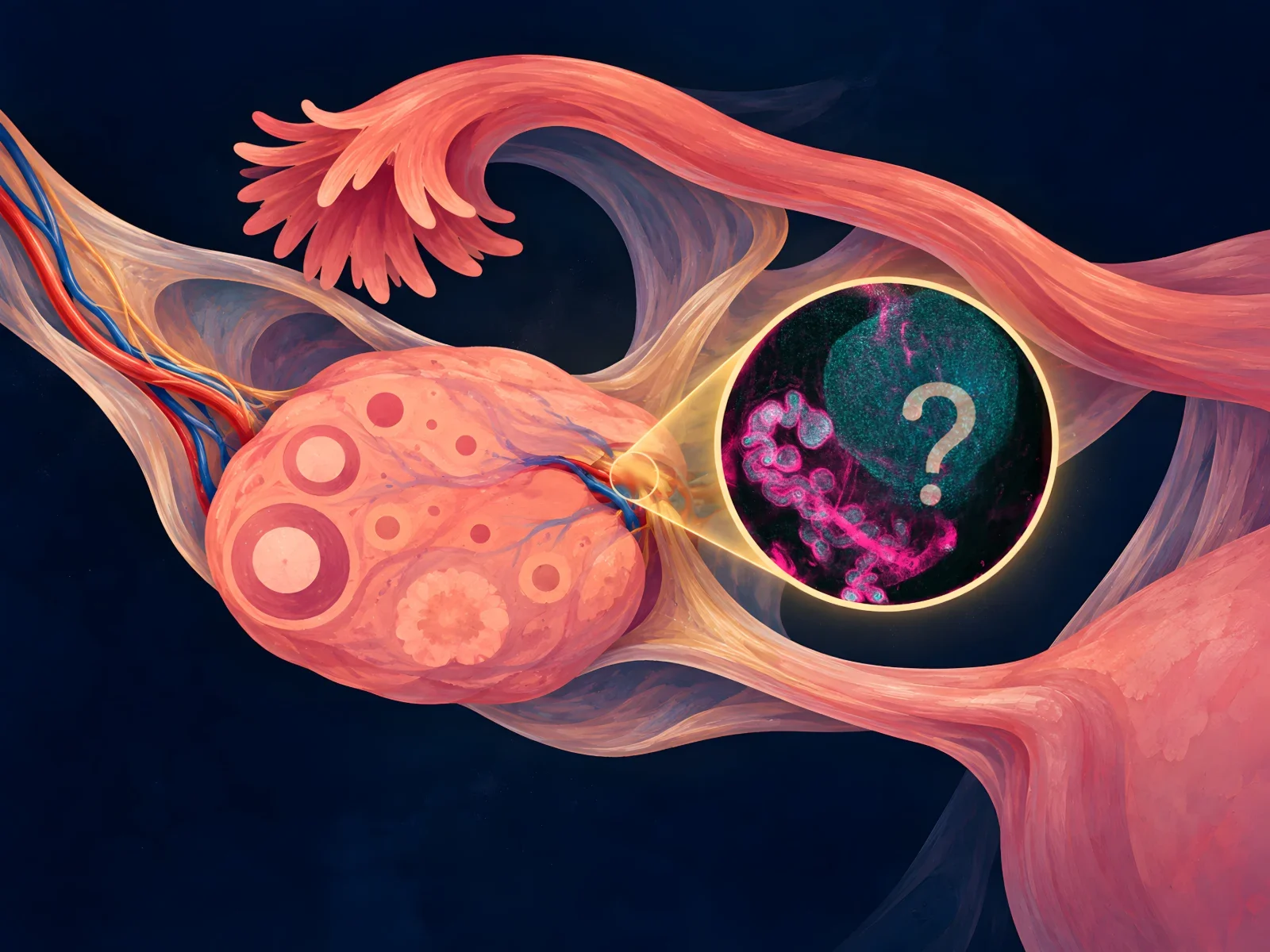
Long recognised in anatomical literature but poorly characterised physiologically, the rete ovarii is now being investigated as a possible sensory and secretory companion to the ovary. What do the experiments actually show, and what might they mean for reproductive medicine?
In 1870, German anatomist Wilhelm Waldeyer described a small epithelial network associated with the ovary. It later appeared in anatomical literature and older illustrations, but its function resisted explanation. The rete ovarii was never truly lost to science. What remained missing was an answer to a surprisingly simple question: why does this network exist alongside an organ whose major functions are described in such detail? [3]
In March 2025, that old question gained new urgency. Researchers studying mouse ovaries introduced a fluorescent tracer into part of the rete ovarii and watched it move towards the ovary. They also found candidate secretory proteins within the structure and close associations with blood vessels, macrophages and neuronal projections. The findings raised a striking possibility: could a structure long regarded as vestigial participate in ovarian signalling—and might that matter for fertility? [1]
What drew me to this research was not the idea of announcing another fertility breakthrough. It was the distance between an anatomical label and a biological explanation. Earlier investigators had questioned whether the rete ovarii was truly functionless. New experimental methods now give that question a more precise form. To understand what has changed, we need to examine its anatomy, development and experimental evidence before considering what the findings may mean for humans.
Understanding the rete ovarii
The rete ovarii is a network of epithelial tubules and cellular cords associated with the ovary, particularly around the ovarian hilum. It develops in close relationship with the embryonic gonad and adjacent mesonephric structures, and has traditionally been considered the female homologue of the rete testis. [2,3]
The rete testis forms an established component of the male reproductive tract, connecting the seminiferous tubules with the efferent ductules and facilitating sperm transport. The rete ovarii, despite its developmental relationship, has no equivalently established function in transporting gametes.
Modern investigations in mice distinguish three anatomically connected regions. The intraovarian rete (IOR) occupies the ovarian medulla and consists predominantly of branching cellular cords. The connecting rete (CR) forms a transitional segment, while the extraovarian rete (EOR) extends beyond the ovary as a convoluted epithelial tube terminating in a dilated, blind-ended structure.
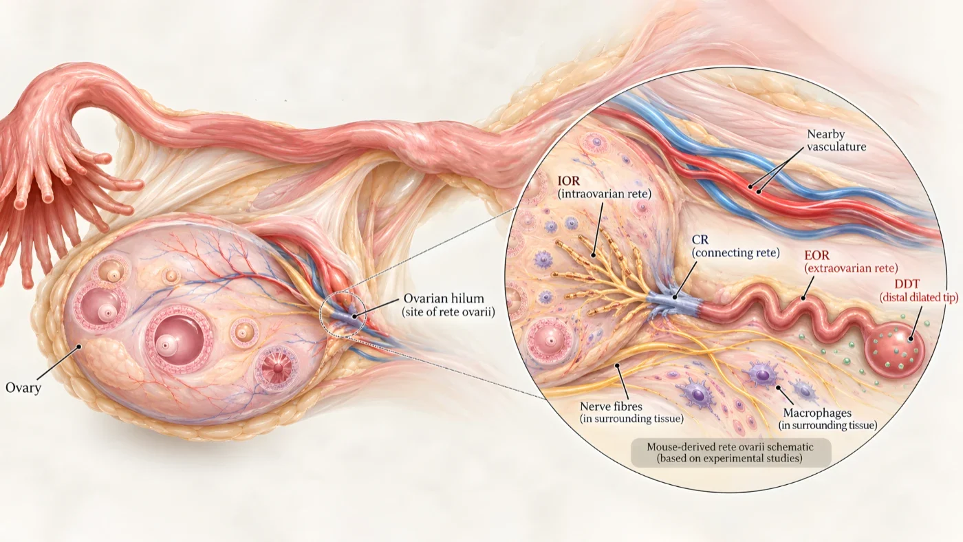
The regions differ in epithelial morphology, molecular-marker expression and developmental behaviour, suggesting that the rete ovarii should not necessarily be understood as a single, functionally uniform structure. [1,2]
The extraovarian portion is particularly interesting because its epithelial cells exhibit features associated with secretory activity. It is also surrounded by vascular structures, macrophages and neuronal projections, placing it in close contact with systems that contribute to ovarian homeostasis.
Developmental regression and the vestigial interpretation
An important observation from the developmental literature concerns how the different regions change as the ovary matures.
In the 2025 study, researchers followed rete ovarii development in mice from embryonic day 16.5 to postnatal day 7.
At E16.5, the connecting and intraovarian regions were relatively prominent. By E18.5, the intraovarian rete had begun to regress, while the extraovarian rete expanded and developed a more conspicuous tubular architecture. At birth, the intraovarian component had become restricted to the ovarian medulla, whereas the extraovarian region remained prominent. [1]
This is a distinction worth emphasising: regression of one anatomical component does not imply disappearance of the entire structure.
Indeed, the same study describes persistence of all three rete regions into adulthood in mice.
Earlier reports of developmental regression helped sustain the interpretation of the rete ovarii as a vestigial structure, although later anatomical investigations questioned whether this classification adequately represented its biology.
Here the meaning of ‘vestigial’ matters. The thymus offers a compelling parallel: once regarded as a vestigial organ, it was later recognised as essential for T-cell development and immune function, demonstrating how a structure’s importance can remain unrecognised long after its discovery. [13]
An evolutionarily reduced structure need not be biologically silent: it may alter an organ’s original role, diminish its contribution or leave it with secondary functions that differ from those of its developmental homologue.
Why did the rete ovarii lose its place in medical teaching?
The rete ovarii did not disappear from scientific research. After Waldeyer’s nineteenth-century description, Byskov and Lintern-Moore investigated its possible contribution to follicle formation in mice in 1973; Wenzel and Odend’hal challenged its classification as a functionless vestige in 1985; and Khan and colleagues examined hormone-receptor expression in human tissue in 1999. None of these studies established a definitive physiological function, but the question had clearly remained open. [3–5]
The 2025 investigators noted that the structure appeared in older Gray’s Anatomy illustrations but was absent from more recent textbook representations. [1] Its uncertain function, limited established clinical relevance and the difficulty of reconstructing its three-dimensional architecture from conventional histological sections offer plausible reasons for reduced prominence in teaching. These are explanations, not documented decisions by individual textbook editors, medical institutions or fertility companies.
The broader history of gaps in women’s health research provides relevant context, although it cannot by itself explain the fate of this particular structure. [12] The more defensible story is not one of a hidden organ suddenly uncovered, but of a familiar anatomical finding whose function remained out of reach until research methods changed.
What the recent research actually demonstrated
A major methodological advance occurred in 2022, when McKey and colleagues applied tissue-clearing techniques and three-dimensional microscopy to study mouse ovarian development in relation to surrounding reproductive structures. Their work demonstrated developmental associations between ovarian morphogenesis, neighbouring tissues and the rete ovarii. [2]
In 2024, Anbarci and colleagues expanded the molecular investigation through transcriptomic analyses of fetal and adult mouse rete ovarii tissues, identifying distinct cellular populations and patterns of gene expression. [6]
These investigations provided the anatomical and molecular framework for the 2025 eLife study, which examined the rete ovarii’s potential secretory function. [1,9]
Evidence of fluid movement toward the ovary
One of the study’s most significant experiments involved introducing fluorescently labelled dextran into the dilated distal tip of the extraovarian rete in postnatal day 7 mouse ovarian tissue. Within approximately 15 minutes, the tracer was detected within the ovary. When the investigators reversed the experiment and introduced dextran near the intraovarian rete, they did not observe equivalent movement into the extraovarian region. [1]
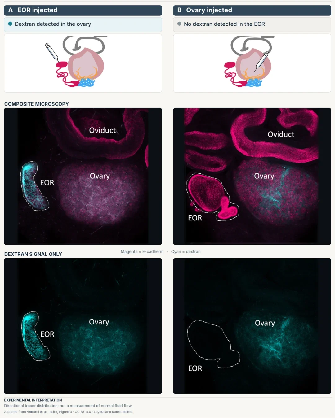
The contrast was revealing: material introduced into the extraovarian rete could reach the ovary under the experimental conditions, whereas the reverse injection did not produce equivalent movement towards the extraovarian region.
This was an experimental demonstration of directional tracer distribution, not a measurement of ordinary fluid flow in a living adult ovary. The distinction matters because it tells researchers exactly what mechanism still needs to be investigated.
The team therefore looked for structures that might help explain the observed movement. The extraovarian rete contains ciliated epithelial cells and is surrounded by tissue expressing smooth-muscle markers. By postnatal day 7, this surrounding layer also expressed calponin, a protein associated with smooth-muscle contractility.
Primary cilia may participate in mechanosensory signalling, while contractile activity could potentially contribute to fluid movement. However, neither mechanism was directly established as responsible for the observed tracer distribution. [1]
The 15 proteins and their biological significance
The tracer experiment raised a second question: what, exactly, might the extraovarian rete carry? Researchers collected luminal contents from postnatal mouse tissue and analysed the samples using mass spectrometry. The analysis detected more than 4,200 proteins. To identify proteins more likely to be associated with secretion, the investigators filtered the results against a mammalian secretome database and compared them with rete-cell transcriptomic data.
Filtering the results in this way narrowed the analysis to 15 candidate secretory proteins. [1]
The significance of this finding has sometimes been exaggerated in online discussions, particularly through claims that the proteins can activate dormant follicles or accelerate egg maturation. Neither effect was demonstrated in the study.
Among these, the investigators examined clusterin (CLU) and syntaxin-binding protein 2 (STXBP2), which are associated with protein handling and vesicle-mediated secretion.
The detection of additional secretory machinery, including components of the SNARE system, strengthened the hypothesis that the extraovarian rete possesses the cellular apparatus required for protein secretion.
One candidate led to an especially interesting line of reasoning: insulin-like growth factor binding protein 2 (IGFBP2). IGFBP2 regulates the bioavailability of insulin-like growth factors, including IGF-1, which participates in granulosa-cell proliferation, steroidogenesis and follicular growth. By binding and sequestering IGF-1, IGFBP2 can reduce the growth factor available for IGF1R-mediated signalling.
The researchers observed relatively high Igfbp2 expression in the extraovarian rete during late fetal development, with lower expression by postnatal day 7. They also identified variation in adult rete-cell expression across stages of the mouse oestrous cycle. [1,6]
From these observations, the investigators proposed a possible feedback mechanism: during oestrus, elevated oestradiol might stimulate rete-derived IGFBP2, which could sequester free IGF-1 and restrain, rather than accelerate, follicular growth and subsequent oestradiol production. [1] This is almost the opposite of the popular suggestion that the 15 proteins have been shown to ‘wake up’ dormant eggs.
I find this proposed feedback loop compelling because it is precise enough to test. Rather than assuming that a neglected structure must encourage fertility, it asks whether rete-derived IGFBP2 could act as a brake within an established growth-factor pathway. The experiment did not test that feedback loop directly; its value is that it now gives investigators a concrete mechanism and predictions to examine.
Could the Rete Ovarii Be the Ovary’s Biological Antenna?
The protein findings suggest one possible output from the rete ovarii. An equally intriguing question concerns its inputs: could this small network sense signals in the surrounding ovarian environment and respond to them? The investigators described this possibility using the metaphor of a ‘biological antenna’. [1]
That metaphor becomes easier to understand when the extraovarian rete is considered in relation to its neighbours. Immunofluorescence studies demonstrated a dense vascular network around the structure, direct contact by neuronal projections, and an association with macrophage populations. [1]
Those neighbours are biologically active, and they suggest several routes by which information could reach rete cells. Its vascular proximity could expose rete cells to circulating hormonal and metabolic signals. Neuronal contacts might influence epithelial behaviour or the activity of surrounding contractile tissue. Macrophages could contribute to local immune regulation, tissue remodelling or inflammatory signalling.
Taken together, these findings sketch a possible system in which the rete receives information about its hormonal, metabolic or local immune environment and changes what it releases towards the ovary. That would make it part of a much wider conversation between ovarian tissues.
For now, it is a model rather than a demonstrated circuit: contact with nerves or vessels and expression of receptors do not, by themselves, show that the ovary changes in response.
But this is precisely why the ‘antenna’ idea is interesting. It turns a striking anatomical arrangement into a set of physiological questions that experiments can answer.
What Do We Know About the Human Rete Ovarii?
The rete ovarii is not exclusive to mice. It has been documented in humans through anatomical, developmental and histopathological investigations.
In 1999, Khan and colleagues examined cellular proliferation, oestrogen and progesterone receptors in human rete tissue. [5]
In 2024, Taelman, Czukiewska and colleagues identified molecular similarities between the fetal rete ovarii and rete testis. [14]
More recently, Lardenois and colleagues analysed approximately 128,000 cells from developing human gonads and linked PAX8-positive rete cells to early supporting-cell lineages. [7]
Together, the human studies extend the story beyond comparative mouse anatomy: they identify rete-associated cell populations during development and suggest a more complicated lineage history than a simple ‘leftover’ description conveys.
Human pathology adds a different kind of relevance. In 1988, Rutgers and Scully described cysts and rare epithelial tumours arising from the rete ovarii, demonstrating its relevance in selected gynaecological pathological diagnoses. [8]
A 2025 preprint proposed another possible role: in genetically altered experimental models, rete ovarii epithelial cells could be transformed into tumours resembling high-grade serous carcinoma. The work is not peer reviewed and does not establish the rete ovarii as a demonstrated origin of these cancers in humans; the fallopian tube remains the principal evidence-supported origin. [11]
These human findings justify studying the structure seriously, while leaving open the question that first drew researchers back to it: what, if anything, does it contribute to normal adult ovarian physiology?
Could the Rete Ovarii Matter for Fertility?
It is difficult to discuss an ovarian structure without eventually asking about fertility. Even small changes in follicular recruitment, hormonal responses or the ovarian environment can matter. Ovarian function depends on coordinated communication among oocytes, granulosa and theca cells, stromal tissue, blood vessels and immune cells. Alongside gonadotropins, local growth factors and paracrine signals help regulate this delicate balance. If rete-derived proteins participate in that network, they could open another avenue for understanding reproductive disorders.
Polyendocrine metabolic ovarian syndrome (PMOS)—the new consensus name for polycystic ovary syndrome (PCOS), with a phased transition beginning in 2026—involves disturbances in ovulation, often accompanied by insulin resistance and hyperandrogenism. [15] Because rete cells express receptors associated with insulin and IGF signalling, researchers could investigate whether their activity changes under altered metabolic conditions. It remains an unproven connection, but one with a specific biological basis for investigation.
Diminished ovarian reserve raises another question: could signals near the ovarian hilum influence follicle maintenance or recruitment? The distinction matters, because recruiting more follicles does not create new ones; accelerated recruitment could instead hasten depletion of the remaining pool. Premature ovarian insufficiency, reproductive ageing and menopause invite related questions about how local ovarian signalling interacts with hormonal and immune changes.
What makes these possibilities worth exploring is the chance to better understand the complex environment that supports ovarian function. Establishing whether rete-cell activity actually influences that environment, and whether the findings extend to humans, would be an important next step. The value of this research lies not in promising another fertility treatment, but in investigating a part of ovarian biology whose contribution we have yet to understand.
What Would It Take to Prove the Rete Ovarii Matters?
The decisive experiment is no longer simply about identifying more proteins or anatomical connections. It is about determining whether changing rete activity changes ovarian function. Do rete-derived molecules reach ovarian cells at physiologically meaningful concentrations? Does secretion respond to hormonal or metabolic changes? And what happens to follicular development when rete-cell activity is selectively altered?
That final experiment is particularly challenging. In the public peer-review record of the 2025 study, investigators described the difficulty of disabling the rete without affecting neighbouring reproductive tissues. The discussion also highlighted the distinction between observing tracer movement and measuring fluid dynamics, and between identifying neuronal contact and demonstrating neural control. [10]
These are not dead ends; they define the next experiments. Demonstrating a reproducible ovarian response, and eventually investigating whether comparable mechanisms exist in humans, would bring us considerably closer to understanding the rete ovarii’s physiological role.
Why I Think This Question Is Worth Pursuing
The rete ovarii is an unusual reminder that anatomy and physiology do not always advance together. We knew what to call this network before we understood how to test its activity. Today, we can describe its changing architecture, trace material from one region towards the ovary and identify candidate signals that may be part of a larger exchange.
What I find most compelling is the shift from asking whether a forgotten-looking structure must be useless to asking what the ovary might gain from its presence. Does it sense local changes? Do its secretions affect follicles? Could it matter more during development than adulthood—or act as a small, supplementary pathway throughout life? Those are sharper questions than the old label allowed.
Fertility research carries real human weight. For people living with the uncertainty of infertility, even modest biological discoveries can feel important. That is exactly why I would rather follow this line of evidence carefully than attach a promise to it too soon. Hope is not strengthened by exaggeration; it is strengthened when researchers ask a question they can genuinely answer.
I don’t need the rete ovarii to become the next major fertility discovery to consider it worth studying. If it turns out to influence ovarian function, our account of reproductive physiology will become more complete. If its contribution is limited, we will have learned something equally important about what the ovary does—and does not—depend on.
For more than 150 years, we have known that the rete ovarii exists. What if the ovary has been responding to it all along?